

EQUIP study: whole-body PET scan from one minute of data
At the EANM congress in Vienna, the EQUIP study of 24 patients rated images rebuilt from the first minute of data at 4.5 out of 5, against 3.9 for a standard scanner.
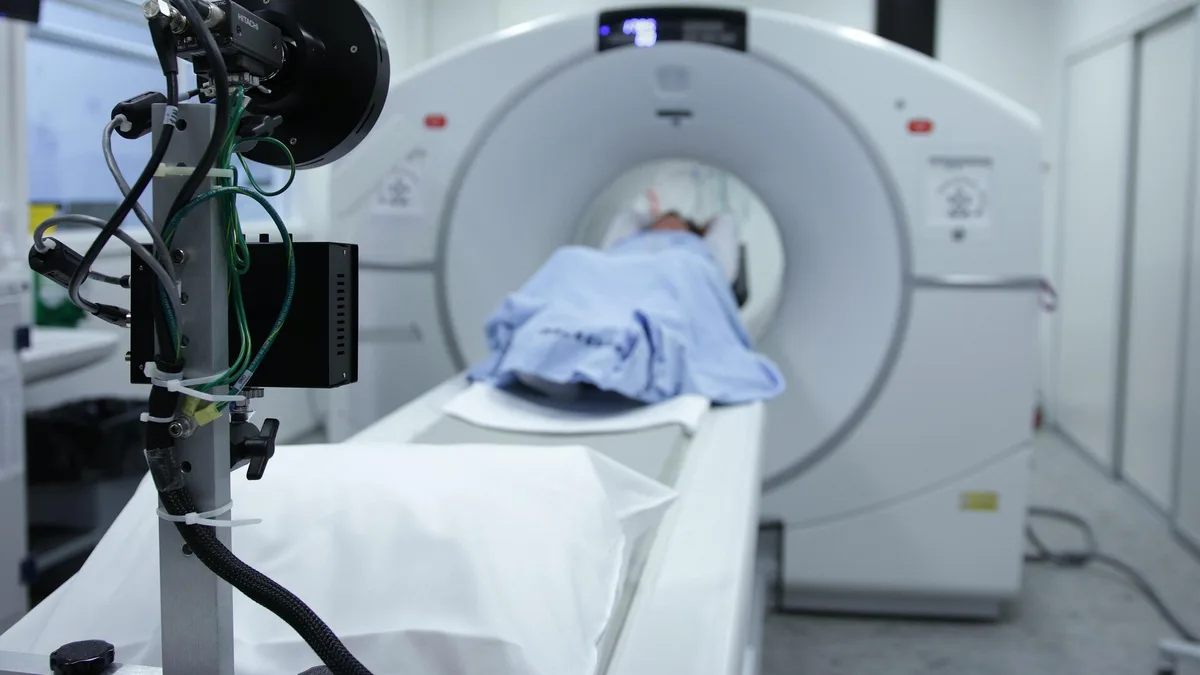
Researchers from Peter MacCallum Cancer Centre in Melbourne presented the results of the EQUIP study on Sunday, October 18, 2026, at the 39th congress of the European Association of Nuclear Medicine (EANM’26) in Vienna, in talk OP-031. Two specialists rated whole-body PET/CT images, reconstructed from just one minute of data, higher than images from the standard scanner used in everyday practice.
What was tested
PET/CT is used to detect, stage and monitor cancer. A conventional scanner has a short field of view, so it images the body piece by piece: from head to thigh takes five to six table positions. Long axial field of view (LA-FOV) scanners cover much more of the body at once and capture more of the signal emitted by the radiotracer, which opens the door to far shorter scans.
The study tested the 128 cm GE HealthCare Omni. The lead author is Dr Cameron Pain, a nuclear medicine physicist with more than ten years of clinical experience. The study is registered on ClinicalTrials.gov as NCT07263815.
How was the comparison made?
Twenty-four patients took part, and each received one of five radiotracers:
- 18F-FDG
- 18F-DCFPyL
- 68Ga-DOTATATE
- 68Ga-PSMA
- 124I
All were first scanned on a standard scanner, then for ten minutes on the new machine. From that data the researchers later reconstructed images using only the first 5, 3, 2 and 1 minutes. In other words, no patient lay in the scanner for only a minute: the shorter images were built from part of the ten-minute scan.
Two nuclear medicine specialists, who did not know how long each scan had taken, scored the images from 1 to 5 on overall diagnostic quality, sharpness, lesion visibility and image noise.
What the scores showed
Average diagnostic quality was 5.0 for the 10-minute and 5-minute images, 4.9 for the 3-minute and 2-minute ones, and 4.5 for the one-minute images. The standard scanner scored 3.9. Every image of two minutes or longer scored at least 4 in every category, and every one-minute image scored at least 3. Image noise showed the largest drop as scan time shortened. The signal-to-noise ratio in healthy liver tissue stayed roughly the same between the 10-minute image and the one-minute one.
Dr Pain said in the EANM statement:
“A one-minute scan on this system gave us images that our readers rated higher than the standard scans we do every day. That was a striking finding: even when we cut the scan time by 90 percent, image quality was above current standard methods.”
The statement lists three possible benefits: less motion from patients who are in pain or find it hard to lie still, more scans per day at a single site, and a lower radiotracer dose. Dr Pain added:
“Total-body PET gives us choices we did not have before. We can use its sensitivity to scan patients faster, to reduce the radiation dose, and to see disease more clearly.”
What these results do not prove
The sample is small, 24 patients, and quality was measured by the scores of two readers, not by diagnostic accuracy. The study therefore does not show that very short scans do not miss small or faint lesions. The authors say larger studies are needed to check that, as well as the effect of faster scanning on workflow and on the patient experience in routine clinical practice.


